Why have I been asked to have a gynaecological ultrasound?
A gynaecological ultrasound is performed in order to assess the anatomy of the uterus, ovaries and other pelvic structures. This can be done in a number of situations:
- To look for causes of pelvic pain.
- To look for causes of problems with periods (heavy periods; periods occurring too frequently; periods occurring irregularly; bleeding between the periods).
- To look for possible causes for difficulties falling pregnant (infertility).
- To look for possible causes for recurrent miscarriages.
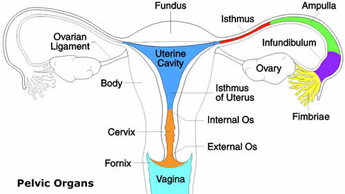
A gynaecological ultrasound is able to provide views of the uterus and ovaries. The fallopian tubes are not usually visible, but may be visible to ultrasound if they are swollen due to a problem. Ultrasound is not good at seeing bowel and therefore a gynaecological ultrasound would not be expected to detect bowel problems.
Do I need a full bladder for my ultrasound?
A full bladder is not required for most ultrasounds. A small amount of fluid in the bladder is often helpful; 1 or 2 glasses of water drunk in the hour before the appointment is usually sufficient.
Will I need a vaginal ultrasound?
In many gynaecological and early pregnancy scans more detailed images may be obtained by a vaginal ultrasound. A vaginal ultrasound is usually less uncomfortable than a pap smear test and should not hurt. You are welcome to refuse if you do not wish to have this type of examination. If a vaginal ultrasound is not appropriate for you, please inform us that you will need an abdominal ultrasound and please attend with a full bladder.
Are there any special techniques used at Specialist Women’s Ultrasound to perform my ultrasound?
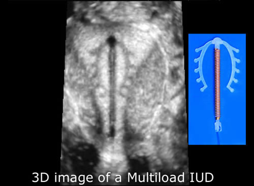
Three-dimensional ultrasound is frequently employed during transvaginal ultrasound to provide clearer images of the uterus and pelvic structures. It enables the shape of the uterus to be far more accurately assessed than with traditional 2D vaginal ultrasound.
Dr Bethune has given several lectures at conferences about the use and benefit of this technique for assessing the uterus.